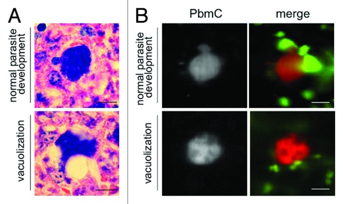
Figure 2. Vacuolization and parasite death is a physiological event that occurs also in vivo. (A) Mice were infected with P. berghei sporozoites and euthanized 38 hpi. The liver was removed, fixed and liver sections were prepared and stained with Giemsa. Parasites developing normally (top panel) are compared with parasites forming vacuoles. Scale bar: 20 µm. (B) Additionally, lys-GFP mice that express GFP mainly in neutrophils and macrophages32 were infected with mCherry-expressing parasites and intravital imaging of the anesthetized mouse was performed between 44 and 48 hpi. At this time parasites developing normally start forming merosomes (top panel, left image; PbmC: P. berghei mCherry), whereas dying parasites form vacuoles (lower panel) and finally fade (see Vid. S3). Scale bar: 20 µm; CLS.
