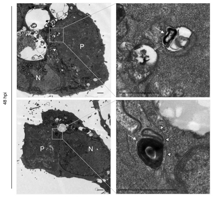
Figure 3. Electron microscopy reveals autophagosome-like structures in P. berghei liver schizonts. P. berghei-infected HepG2 cells were fixed 48 hpi and analyzed by electron microscopy. P, parasite; N, host cell nucleus. Arrows indicate autophagic-like vesicle with multiple membranes, arrowheads indicate the parasite plasma membrane. Boxed areas are displayed at a higher magnification in the right panel. Scale bar: 2 µm.

An official website of the United States government
Here's how you know
Official websites use .gov
A
.gov website belongs to an official
government organization in the United States.
Secure .gov websites use HTTPS
A lock (
) or https:// means you've safely
connected to the .gov website. Share sensitive
information only on official, secure websites.
