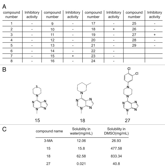
Figure 2. Identification of autophagy inhibitors by imaging-based screening. (A) GFP-LC3-NRK cells were starved for 4 h with or without 3-MA derivatives at 0.1 mM, 1 mM or 10 mM and at least three replicates were performed. Cells were observed by confocal microscopy and formation of GFP-LC3 puncta was assessed by visual screening. Activity of the compounds was scored as “+” if the formation of starvation-induced puncta was inhibited by the addition of the respective compound (at least with the two higher doses) and “-” if no inhibition of puncta formation was observed. (B) The structures of the three autophagy inhibitors identified by screening. (C) The solubility of 3-MA and the three active derivatives in DMSO or water at 37°C.

An official website of the United States government
Here's how you know
Official websites use .gov
A
.gov website belongs to an official
government organization in the United States.
Secure .gov websites use HTTPS
A lock (
) or https:// means you've safely
connected to the .gov website. Share sensitive
information only on official, secure websites.
