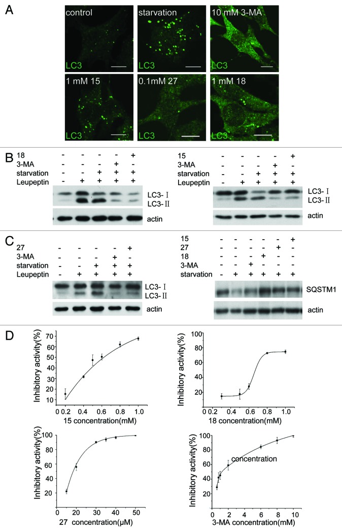
Figure 3. Derivatives 15, 18 and 27 inhibit autophagy in NRK cells. (A) NRK cells were starved for 4 h with or without 10 mM 3-MA, 0.1 mM 27, 1 mM 18 or 1 mM 15. Cells were stained with an anti-LC3 antibody and observed by confocal microscopy. Scale bar: 10 μm. (B) Leupeptin (10 μg/ml)-treated NRK cells were starved for 4 h with or without 10 mM 3-MA, 1 mM 18, 1 mM 15 or 0.1 mM 27, and processing of LC3 was measured by western blot using anti-LC3 antibody. (C) NRK cells were starved for 4 h with or without 10 mM 3-MA, 1 mM 18, 1 mM 15 or 0.1 mM 27 and the level of SQSTM1 protein was measured by anti-SQSTM1 antibody. (D) Dose-response of the three compounds compared with 3-MA. GFP-LC3-NRK cells were starved for 4 h with or without the indicated compounds. The level of LC3-positive puncta per cell was quantified in at least 30 cells. Autophagy inhibition index = % (1 − (a − b) / (c − b)), where “a” means the number of GFP-LC3-positive puncta per cell in cells treated with compound + DPBS; “b” means the number of GFP-LC3-positive puncta per cell in DMSO-treated cells; “c” means the number of GFP-LC3-positive puncta per cell in DPBS-treated cells. Results were analyzed with OriginPro8 software.

An official website of the United States government
Here's how you know
Official websites use .gov
A
.gov website belongs to an official
government organization in the United States.
Secure .gov websites use HTTPS
A lock (
) or https:// means you've safely
connected to the .gov website. Share sensitive
information only on official, secure websites.
