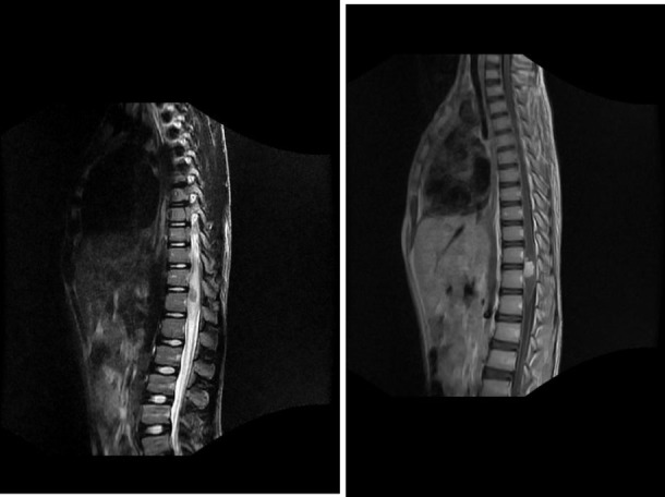
Fig 1.

Sagital T2 (A) (FSE TR3200 TE 110 FOV 36) and T1 post-contrast (B) (FSE TR 625 TE min full FOV 36) images in a 15 year old male with acute onset paraplegia show an enhancing anterior conus lesion (straight arrows) with extensive cord edema (curved arrows). The focality allowed for surgical resection, and histological analysis of the tissue revealed the presence of Schistosoma haematobium eggs.
