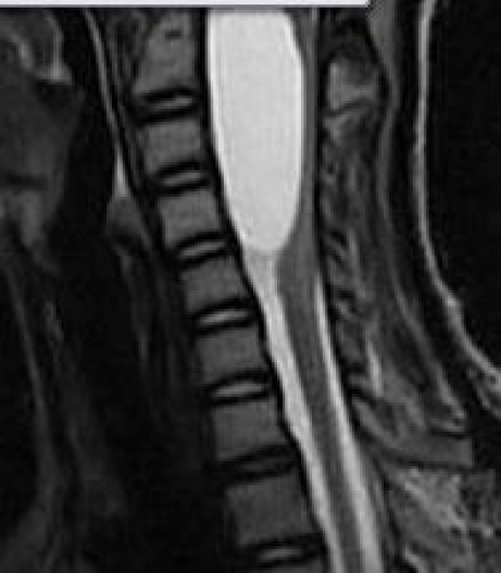
Figure 2.

A sagital T2 (A) (FSE TR3200 TE 110 FOV 36) image of the cervical spine in a 14 year old female with progressive subacute quadriparesis. The image shows an anterior, intradural and extra-medullary cystic mass with homogenous signal intensity consistent with an enlarging arachnoid cyst. There is associated effacement and compression of the cervical cord, with posterior displacement. This image was the basis for surgical planning and resection which led to a successful neurological outcome.
