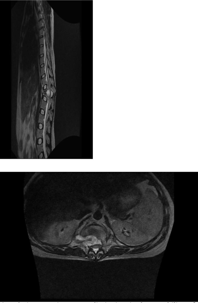
Fig 3.

agital T2 (A) (FSE TR3200 TE 110 pFOV 36) and Axial T2 (B) (FSE TR 3800 TE 108 28 pFOV) images of a 26 year old male who presented to Queen Elizabeth Central Hospital (QECH) following acute onset of paraplegia. MRI imaging allowed for localization and identification of the offending lesion (arrows) as spinal TB, as well as defining its extent, both of which were critical in surgical planning for resection.
