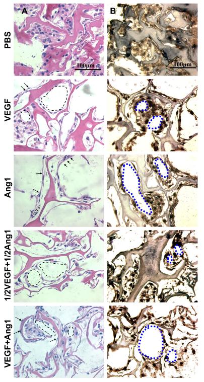
Figure 6. Cell morphology and organization after 7-day in vitro cultivation.
(A) Hematoxylin and eosin stained images (173,333 cells initially seeded on the freshly made scaffolds; arrows indicate elongated cells; arrowheads indicate circular structures). Note that darker pink in hematoxylin and eosin staining indicates the collagen scaffold, while the lighter pink is part of the cells. (B) Von Willebrand factor stained images (173,333 cells initially seeded; brown represents positive von Willebrand factor staining, blue represents counterstain; circular structures indicated by blue dotted outlines). Figure reproduced from [14] with permission. Copyright Elsevier.

