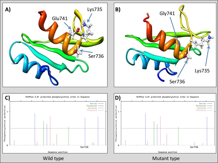
Fig. 2.
Ab-initio 3D modeling of SH2 domain (VAV3). (a) Wild type, note hydrogen bond between Glu741 and Lys735 (blue line); (b) Mutant type, hydrogen bond between Gly741 and Lys735 cannot be formed; (c) and (d) posphorylation potential of serine (blue), threonine (green), and tyrosine (red). Threshold is marked by horizontal gray line (phosphorylation analysis performed by NetPhos 2.0).

