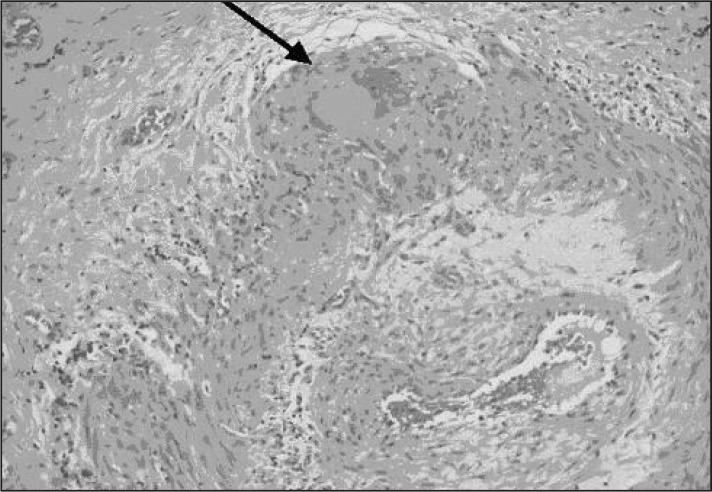
Figure 1.
Histologic specimen of giant cell arteritis. Arrow points to giant cell in arterial wall.
Reprinted with permission from: Mansoor O, Majeed T. A 90 year old woman with painless vision loss. Digital Journal of Ophthalmology [serial on the Internet] 2005 Feb 10 [cited 2012 Oct 31];11(7): Figure 2. Available from: www.djo.harvard.edu/site.php?url=/physicians/gr/728&page=GR_TT.

