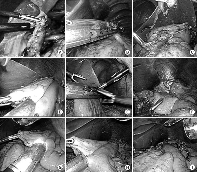
Fig. 3.
Intracoporeal BI anastomosis (A, B, C), intracoporeal BII anastomosis (D, E, F), intracoporeal Roux en Y anastomosis (G, H, I). (A) Cut of duodenum upper edge. (B) Closure of endo GIA entry hole. (C) Laparoscopic view of the gastroduodenostomy. (D) Anastomosis with endo GIA. (E) Closure of endo GIA entry hole. (F) Laparoscopic view of the gastrojejunostomy. (G) Jejunojejunostomy with endo GIA. (H) Gastrojejunostomy with endo GIA. (I) Laparoscopic view of Roux en Y anastomosis.

