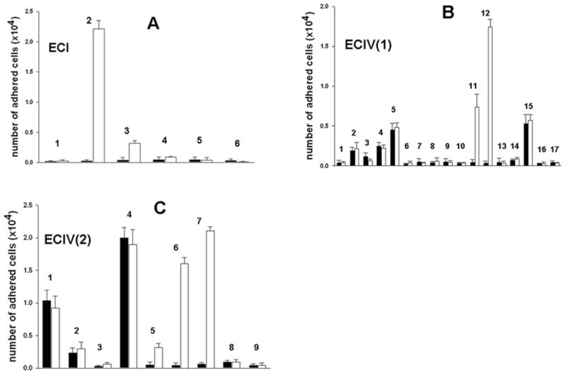
Fig. 3.
Screening of fractions ECI (A), ECIV(1) (B), and ECIV(2) (C) for pro-adhesive properties in the cell adhesion assay. Fractions (1 μg in 0.1 ml of PBS) were immobilized on 96-well plate by overnight incubation at 4°C. K562 cells (filled bars) and α2K562 cells (open bars) were labeled with CMFDA and applied on the wells previously blocked with 1%BSA. After 30 min incubation, unbound cells were removed by washing, whereas adhered cells were lysed with 0.5% Triton X-100. Plate was read with fluorescence plate reader using 485 nm excitation and 530 nm emission filters. Error bars represent S.D. from three independent experiments.

