
Figure 1.
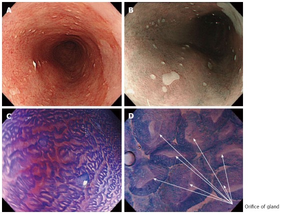
White light endoscopy, narrow-band imaging and endocytoscopy examination of long segment Barrett’s esophagus. A: Long segment Barrett’s esophagus under white light endoscopy (WLE); B: Narrow-band imaging with low magnification clearly visualized small squamous cell islands within regular columnar Barrett's epithelium, which are also identified by WLE with difficulty; C: Endocytoscopy (ECS) examination after crystal violet and methylene blue (CM) double staining; D: ECS examination under higher magnification (× 480) shows the glandular orifices of regular Barrett’s epithelium.