
Fig. 1.
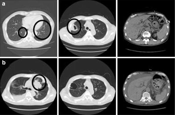
a Contrast-enhanced computed tomography (CT) performed on the eighth hospital day showed small nodular lesions scattered in both lung fields (left), cavity formation in the upper lobe of the right lung (middle), and ascites and slight liver atrophy (right). b CT performed on the 38th hospital day showed a nodular lesion, lung metastases, and massive pleural effusion in the right thoracic cavity (left and middle). Liver atrophy was aggravated compared with the above image (right) (b)