
Fig. 3.
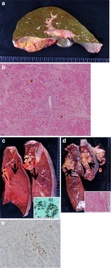
Autopsy findings. a Macroscopic view of the liver shows mild atrophy and marked cholestasis. b H&E staining of the liver shows massive liver necrosis and moderate lymphocyte infiltration, and cholestasis (magnification 10×). c Congested lung and marked overgrowth of Aspergillus mycelia in the bilateral lungs. Arrows show overgrowth of Aspergillus mycelia. Lower right box shows Grocott’s methenamine silver staining of Aspergillus mycelia in these lesions. d Metastatic lesions in the bilateral lungs. Arrows show these lesions. Lower right box shows H&E staining of metastatic renal carcinoma in these lesions. e HBs-Ag immunostaining in the liver