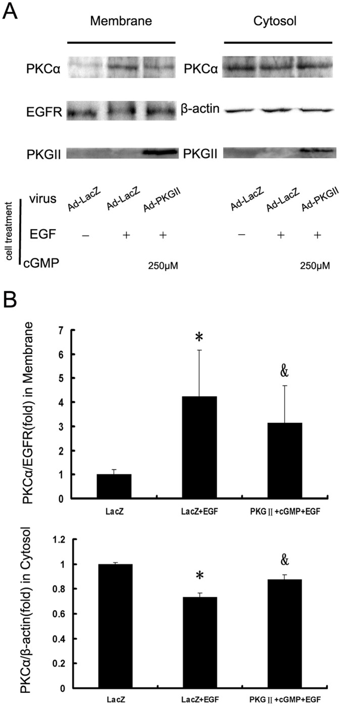
Figure 7. PKG II prevents the activation of PKCα.
AGS cells were treated same as described in Figure 4. Subcellular fractionation into cytosol and membrane fractions was performed by using Membrane and Cytosol Protein Extraction Kit. Western blotting was used to detect PKCα either on the membrane or in the cytosol. Densitometry analysis was performed to quantify the positive bands. A: A representative of initial results of three independent experiments. B: Results of densitometry analysis. The data shown are the means ± SD from 3 independent experiments (*P<0.05, compared to LacZ group; &P<0.05, compared to LacZ+EGF group).

