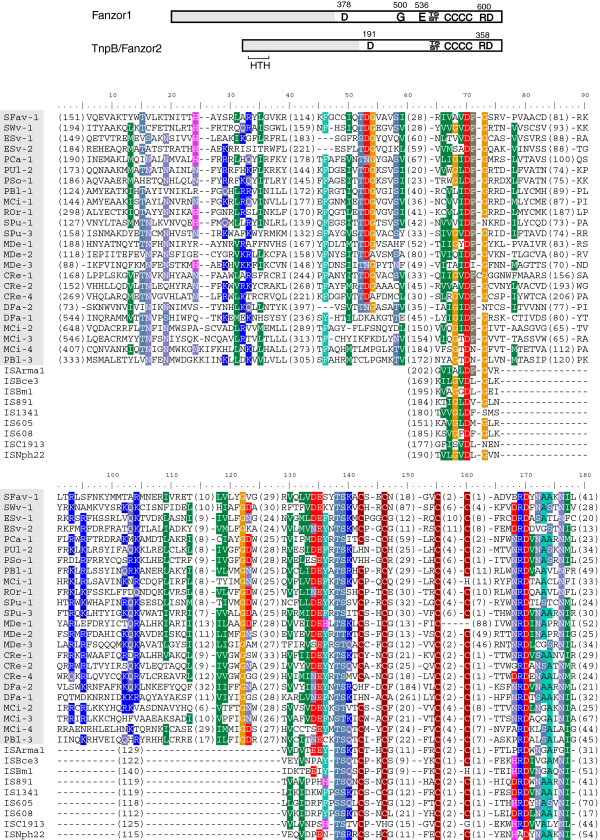
Figure 1.
Motifs and alignments of Fanzor and TnpB proteins. Conserved amino acids and helix-turn-helix (HTH) domain are marked (above); gray regions indicate the variable N-terminal halves. Numbers above the diagram refer to the residue position in SPu-1-1p or TnpB_IS608. Titles of Fanzor1 proteins are shaded in the alignment.

