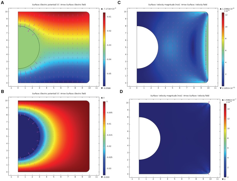
Figure 1. Electric field and electroosmotic velocity in the cylindrical model of the polarized and non-polarized cells.
Results of the computer simulation of electric potential, electric field and electroosmotic flow velocity in the axisymmetric model of the cell (cylinder of 10 µm radius and 10 µm height, nucleus–sphere of 3 µm radius, located in the center of the cylinder). Mathematical model is described by eqs. (1–6); parameters are presented in Table 1. Figure 1A represents electric potential (color map) and electric field strength (arrows) in the model of the polarized cell; inward current enters through the top of the cylinder and leaves the cell through the bottom of the cylinder (eq. 2). Electric potential is in the range −0.007÷−0.059 V; strong component of electric field parallel to the cylindrical surface is present. For comparison, Figure 1B presents electric potential and field in the non-polarized cell (zero inward and outward current); here the component of electric field parallel to the surface is practically absent. Figure 1C demonstrates electroosmotic flow circulation (color represents magnitude of the velocity, arrows represent direction) caused by electric field distribution in the polarized cell (Fig. 1A), maximum value of fluid velocity 120 µm/s is in the vicinity of the cylindrical wall, while the average magnitude of the fluid velocity across the cytoplasm is 20 µm/s. Fluid circulates, moving downwards near the cylindrical wall and upwards near the nucleus. For comparison, Figure 1D demonstrates that electroosmotic flow is practically absent (except the filleted corners) in the model of a non-polarized cylindrical cell, where electric field parallel to the surface is absent.

