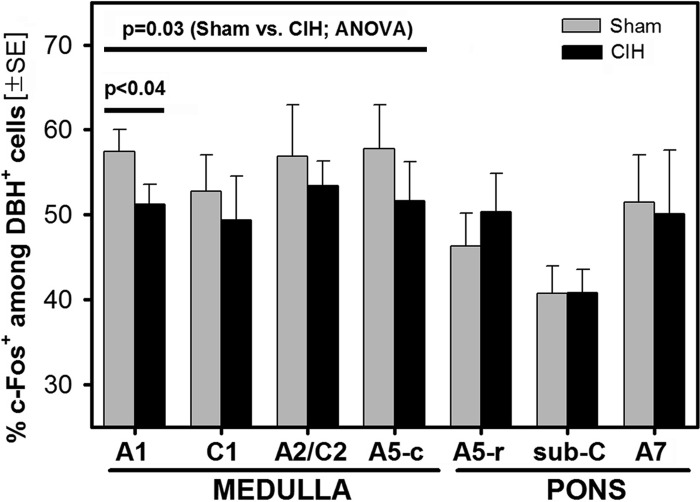
Fig. 4.
Percentages of DBH cells that had c-Fos-stained nuclei in different groups of pontomedullary catecholaminergic neurons. The percentage of DBH-positive (DBH+) cells that were c-Fos-positive (c-Fos+) was significantly lower, or tended to be lower, in the CIH- than sham-treated rats in all medullary groups [A1, C1, A2/C2, and caudal A5 (A5-c); significant effect of the treatment by two-way ANOVA, 6 rats per treatment group]. The effect was also significant for the A1 group when tested by a paired t-test. In contrast, in the pons [rostral A5 (A5-r), sub-C and A7], CIH, and sham animals did not differ. All animals were perfused at the same time of the day 1 day after 35 days of either daily CIH exposure or sham treatment and with wakefulness maintained before perfusion by exposure to a novel environment.

