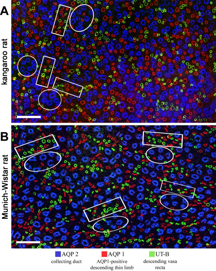
Fig. 4.

Immunolocalization of tubules and vessels in the upper inner medulla. A: kangaroo rat; B: Munich-Wistar rat. Intracluster regions (circles) consist predominantly of CDs. Intercluster regions (rectangles) consist of DTLs and DVR. Interconnecting capillaries and ATLs of the intracluster region and ATLs and nonbranching AVR of the intercluster region are not shown. Both images are overlays of two sections no more than 3 μm apart. Transverse sections are from ∼900 μm below the outer medulla. Scale bars: 100 μm. Figure was modified from Issaian et al. (48).
