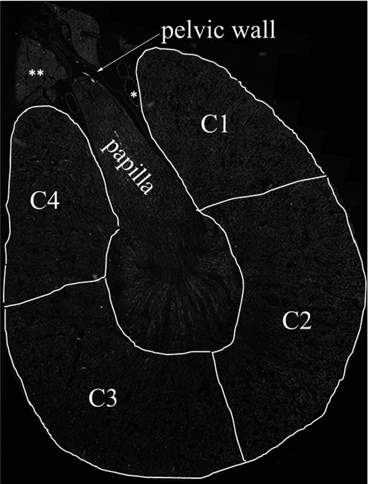
Fig. 1.
For analyses of the optical intensity of the renal sensory nerves, the renal pelvic wall was outlined (arrow) and for analyses of the intensity of the sympathetic nerves four areas of the renal cortex were outlined, C1 through C4. C1 and C4 represent the peripelvic area, and C2 and C3 represent the renal cortical areas more distal from the renal hilus. C2 and C3 also included the outer part of the outer medulla. *Vessels; **fat tissue.

