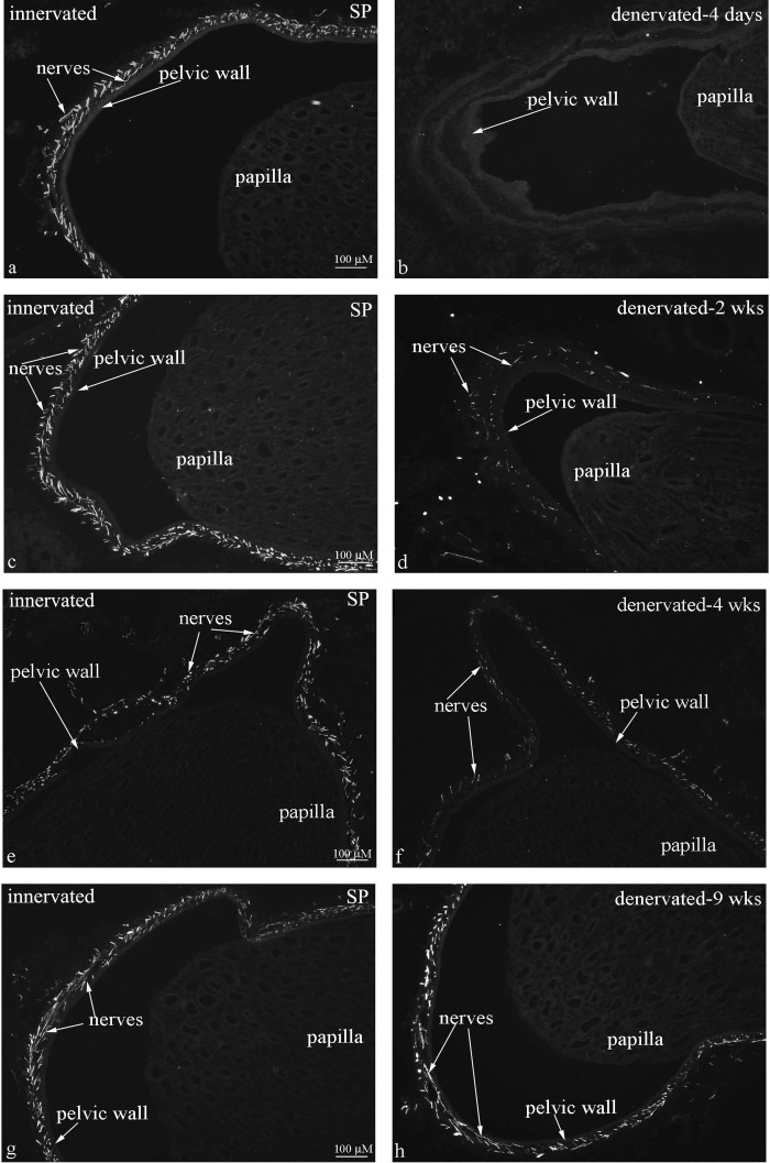
Fig. 4.
Immunohistochemical labeling of renal tissue for substance P (SP) showed dense distribution of SP-ir fibers in the renal pelvic wall in innervated kidneys at all time points (left). In the contralateral denervated kidneys (right), there were no SP-ir fibers in the pelvic wall 4 days after denervation (b) and markedly reduced numbers at 2 and 4 wks after denervation (d, f). At 9 wks after denervation, the distribution of SP-ir fibers in the pelvic wall was similar in the innervated and denervated kidneys (g, h).

