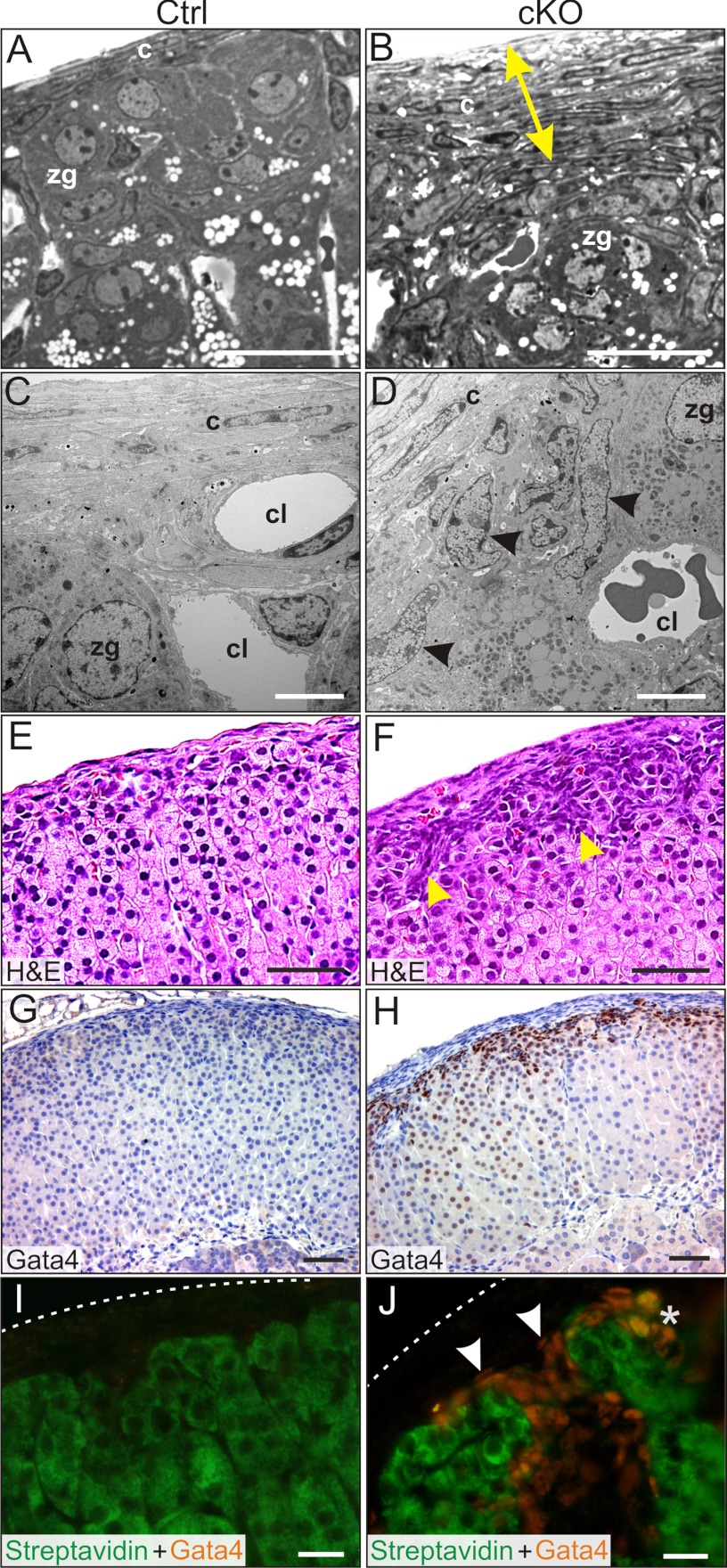
Figure 3.
Capsular and subcapsular changes in the adrenal glands in Gata6 cKO mice. A and B, Semi-thin sections of adrenal gland from 2-month-old control (A) and cKO (B) virgin female mice. Note the thickened capsule in the mutant adrenal gland (yellow arrow). C and D, EM of adrenal glands from 2-month-old control (C) and cKO (D) virgin female mice. Note the accumulation of type A cells (arrowheads) beneath the capsule. E and F, H&E-stained control (G) and cKO (H) adrenal glands from 3-month-old parous female mice. Note the thickened capsule and the accumulation of basophilic type A cells in the subcapsule (arrowheads). G and H, GATA4 immunoperoxidase staining of adrenal glands from 3-month-old control (G) and cKO (H) parous female mice. Note the intense nuclear immunoreactivity in the subcapsule of the cKO adrenal gland. GATA4 immunoreactivity was also evident in some zF cells of the cKO mice. I and J, Adrenal glands from 2-month-old control (I) and cKO (J) virgin female mice stained with FITC-steptavidin and anti-GATA4 (Cy3-labeled secondary antibody). Dashed lines indicate the surface of the capsule. FITC-streptavidin labels steroidogenic and presteroidogenic adrenocortical cells (43). Nonsteroidogenic type A cells exhibit nuclear GATA4 immunoreactivity but do not stain with streptavidin (J, arrowheads). *, Note the rare subcapsular cells that stain with both streptavidin and anti-GATA4 (J) and are surmised to have steroidogenic potential. Abbreviations: c, capsule cell; cl, capillary lumen. Scale bars, 10 μm (A, B, and E–H), 2 μm (C and D), and 30 μm (I and J).

