Figure 5.
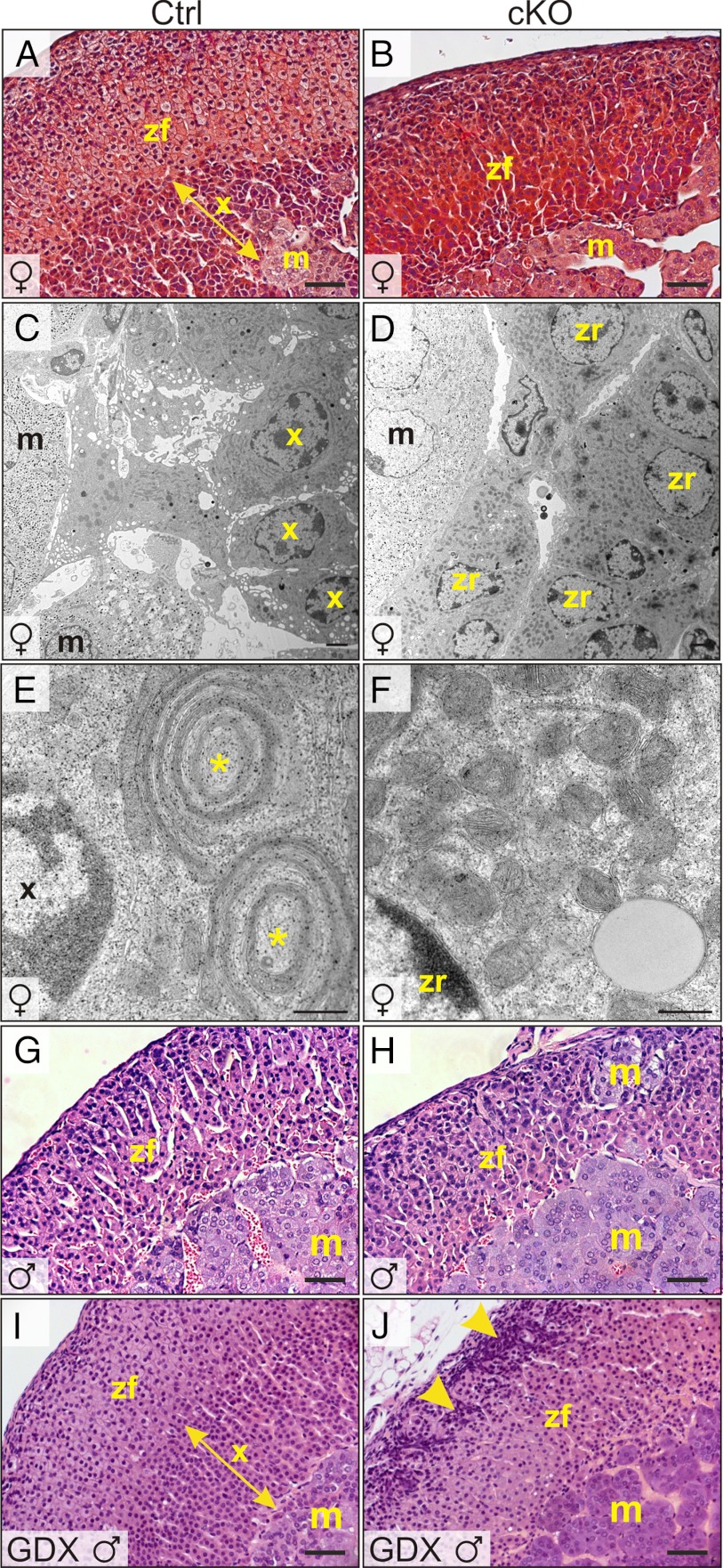
Absence of an adrenal X-zone in Gata6 cKO mice. Adrenal glands from control (A, C, E, G, and I) or cKO mice (B, D, F, H, and J) were processed for light (A, B, G, and H) or EM (C–F). A and B, Trichrome staining of 1-month-old female adrenal glands highlights the lack of an X-zone in the mutant. C–F, Adrenal glands from 2-month-old virgin female control mice contain cells with the ultrastructural features of the X-zone, including dense cytoplasm, abundant smooth ER, and distinctive mitochondrial complexes consisting of flattened mitochondria alternating with ER cisternae (*). The 2-month-old virgin female cKO adrenal glands lack cells with the hallmarks of X-zone; instead, cells with ultrastructural features of zR, including electron-lucent lipid droplets and numerous ellipsoidal mitochondria, abut chromaffin cells in the adrenal medulla. G and H, H&E staining of adrenal glands from 2-month-old control or cKO male mice. I and J, H&E staining of adrenal glands from 2-month-old male mice that were subjected to orchiectomy at 3 weeks of age. Note the secondary X-zone in the orchiectomized control mice. In contrast, the adrenal glands of orchiectomized cKO mice lack an X-zone and have pronounced subcapsular cell hyperplasia (arrowheads). Scale bars, 10 μm (A, B, and G–J), 250 nm (C and D), and 25 nm (E and F). Abbreviations: m, medulla; x, X-zone.

