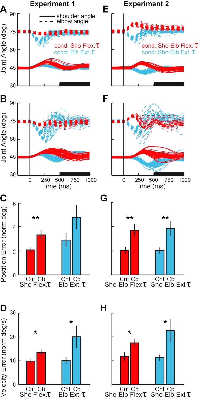
Fig. 3.
Postural stabilization and final accuracy. A: joint motion from an exemplar control subject after the perturbations of experiment 1, shoulder flexion torque (red) and elbow extensor torque (blue). Shoulder and elbow angles of each trial are depicted with solid and dashed lines, respectively. Vertical line shows perturbation onset. B: joint motion from an exemplar subject with cerebellar damage, same format as in A. C: position error (mean and SE) from 500–1,000 ms after perturbation onset (see black bar on time axis in A and B). Red and blue bars correspond to shoulder torque and elbow torque conditions, respectively. Cnt, normal subjects; Cb, cerebellar subjects. D: velocity error (mean and SE) from 500–1,000 ms after perturbation onset, same format as in C. E–H: joint motion of control subjects and cerebellar subjects during experiment 2; same format as in A–D. Significant difference between cerebellar and control groups for the perturbation condition: *P < 0.05; **P < 0.01.

