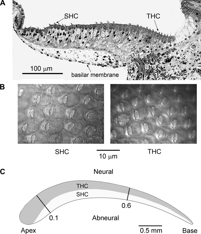
Fig. 1.
Hair cell arrangement in the chicken basilar papilla. A: transverse section through a papilla in the apical region [fractional distance from apex (d) = 0.25] showing the location of the short hair cells (SHCs) above the basilar membrane and the tall hair cells (THCs) overlying the fibrocartilaginous plate containing the auditory nerve fibers; P1 chicken. B: surface views of the intact papilla showing the lower density of the SHCs with their eccentrically placed hair bundles and the higher density of THCs. The low surface density and placement <10 cells from the abneural edge were used to identify SHCs for recording. C: schematic of basilar papilla showing the longitudinal section over which most of the recordings were made from d = 0.1 to 0.6 and the approximate distributions of the THCs and SHCs based upon Hirokawa (1978; Fig. 2b) and the surface morphology of whole mount preparations as in B. In some descriptions, the apical and basal ends of the papilla are referred to as “distal” and “proximal,” respectively, and the neural and abneural edges are termed “superior” and “inferior.”

