Fig. 1.
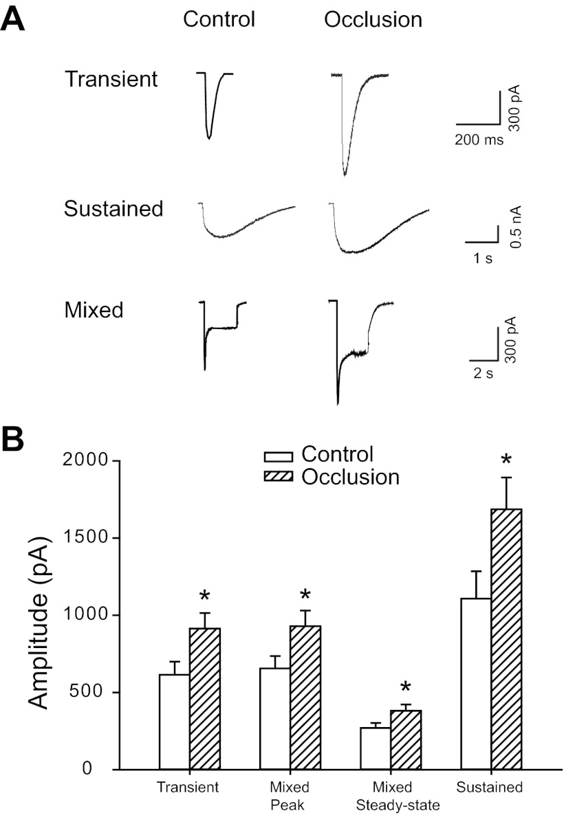
Whole cell patch-clamp methods were employed to study effects of 24-h femoral arterial occlusion on P2X currents induced by α,β-methylene ATP (α,β-meATP). A: original traces showing currents elicited by 30 μM α,β-meATP in dorsal root ganglion (DRG) neurons of control limbs and limbs with femoral artery ligation. Three different P2X currents were observed in both the control and occlusion groups, including the fast transient (top), sustained (middle), and mixed currents (bottom). The amplitudes of the 3 types of currents in occlusion limbs were much larger than those obtained in control limbs. B: averaged data show mean amplitudes of currents activated by α,β-meATP. Twenty-four hours of arterial occlusion significantly increased amplitudes of transient currents, both peak and steady-state of mixed currents, and sustained currents compared with control. *P < 0.05 vs. control.

