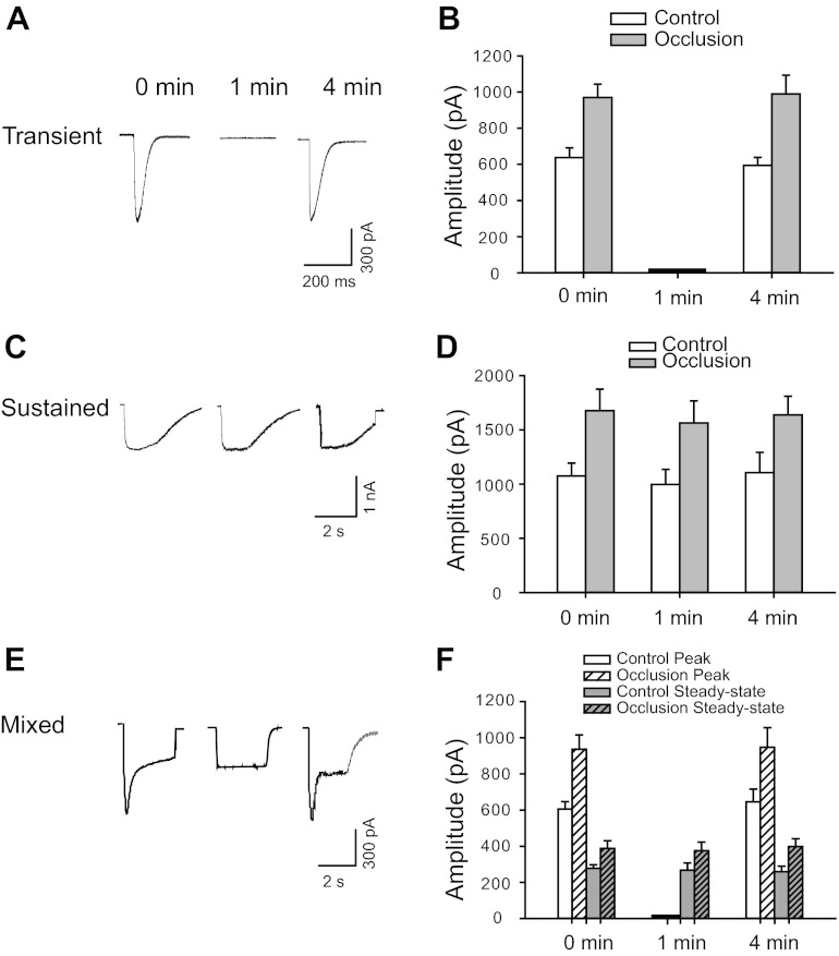
Fig. 3.
Desensitization of ATP-induced currents in DRG neurons innervating muscle. Original traces from control group (A, C, and E) and averaged data (B, D, and F) from control and occlusion groups show currents of DRG neurons activated by application of 30 μM α,β-meATP at different time intervals. A and B: transient current desensitized rapidly. The original peak amplitude was restored at intervals longer than 4 min between consecutive α,β-meATP applications (P = 0.73 in control and P = 0.76 in occlusion between 0 and 4 min). C and D: sustained current was elicited repeatedly and reached its original amplitude at 1-min intervals (P = 0.56 in control and P = 0.63 in occlusion among intervals). E and F: in the cells exhibiting mixed responses, the peak component exhibited desensitization characteristics similar to those of transient currents. The steady-state component of mixed currents demonstrated behavior similar to that of pure sustained currents. Overall, no significant difference was observed in the desensitizing property of the currents elicited by α,β-meATP between control and 24 h of arterial occlusion.

