
Fig. 4.
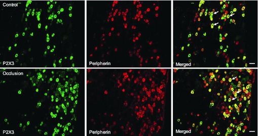
Immunofluorescence was employed to examine double labeling for P2X3 and peripherin. Peripherin was used to label DRG neurons that project thin C-fibers. Representative photomicrographs show P2X3 and peripherin staining in DRG neurons of a control limb (top) and an occluded limb (bottom). Arrows indicate representative cells positive for both P2X3 and peripherin after the images were merged. The number of double-labeled DRG neurons is greater in occluded limbs than in control limbs. Scale bar, 50 μm.