
Fig. 5.
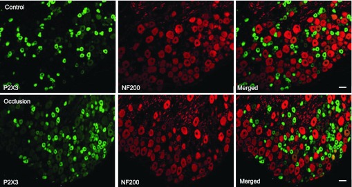
Immunofluorescence was employed to examine double labeling for P2X3 and NF200. NF200 was used to identify A-fibers of DRG neurons. Representative photomicrographs show P2X3 and NF200 staining in DRG neurons of a control limb (top) and an occluded limb (bottom). Very few DRG neurons appeared to be both P2X3 and NF200 positive. No differences in the number of DRG neurons double-stained for P2X3 and NF200 were observed in the control and occluded groups. Scale bar, 50 μm.