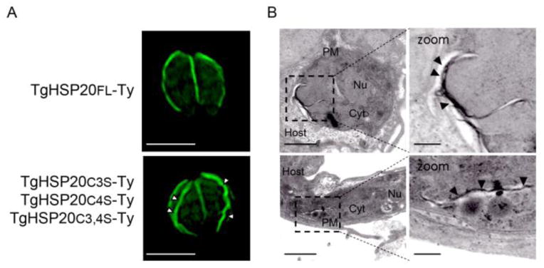
Figure 3. Transmission electron microscopy of the TgHSP20C3,4S-Ty construct suggests the presence membrane accumulation.

Lack or partial palmitoylation at the N-terminal of TgHSP20 forms an unidentified structure. A) Immunofluorescence assay showing the sub-cellular localization of full-length TgHSP20-Ty (TgHSP20FL-Ty; upper panel) or parasites over-expressing different N-terminal palmitoylable TgHSP20 constructs (same result either with TgHSP20C3S-Ty, TgHSP20C4S-Ty or TgHSP20C3,4S-Ty; lower panel). Scale bars= 5 μm. B) Transmission electron microscopy of the parasites observed in panel A. The white arrowheads show the atypical localization of TgHSP20 with indirect immunofluorescence and black arrowheads in the electron microscopy analysis. PM: plasma membrane; Nu: nucleus; Cyt: cytoplasm. Scale bars= 1 μm and 200 nm for the zoomed images.
