Figure 3.
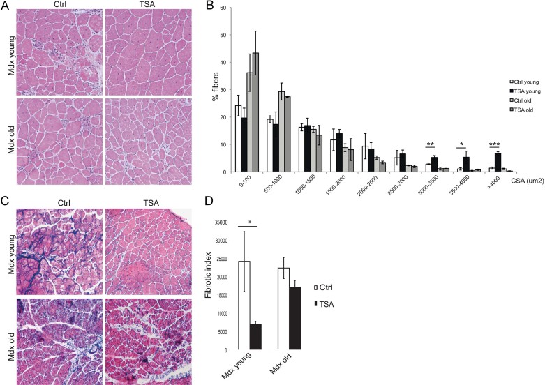
Skeletal muscles from aged MDX mice are resistant to HDACi-induced beneficial effects.
A. Representative images of Hematoxylin and Eosin staining of tibialis anterior transverse sections of young (1.5 months old) and old (1 year old) mdx mice treated for 45 days with vehicle (Ctrl) or TSA (0.5 mg/kg).
B. Graph of the analysis of myofibre cross sectional area (CSA) of muscles represented in (A) showing that TSA increases the caliber of myofibres only in young MDX muscles. Data are represented as average ± SEM. n ≥ 3. Statistical significance assessed by t-test, *p < 0.05, **p < 0.01, ***p < 0.01.
C. Representative images of Masson's trichrome staining of tibialis anterior transverse sections of young (1.5 months old) and old (1 year old) MDX mice treated for 45 days with vehicle (Ctrl) or TSA (0.5 mg/kg).
D. Graph representing the fibrotic index (quantification of collagen deposition) measured as blue area (reported as pixel2) per field. Data are presented as the average ± SEM. n ≥ 3. Statistical significance assessed by t-test, *p < 0.05.

