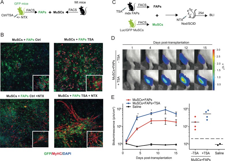
Figure 5.
HDACi enhance the ability of FAPs to promote myogenic activity of MuSCs on injured muscles ex vivo and in vivo.
A. Schematic representation of co-culture strategy. GFP – FAPs were isolated from notexin injured (+NTX) or non-injured (ctrl) skeletal muscles of GFP mice (n = 2), treated with vehicle (ctrl) or TSA for 5 days, and then co-cultivated for 14 days with MuSCs isolated from wt mice.
B. Representative images of immunostaining for MyHC (red) and GFP (green) of co-culture between FAPs from ctrl (left panel) and TSA (right panel) GFP mice and wt MuSCs. Insets show a higher magnification.
C. Schematic representation of MuSCs/FAPs co-transplantation into the irradiated tibialis anterior of immunodeficient Nod/SCID mice. MuSCs were isolated from double-transgenic Luciferase-EGFP mice. FAPs were isolated from mdx mice treated with TSA or control (vehicle) for 5 days. Twenty-four hours prior to intramuscular MuSCs/FAPs co-transplantation, recipient mice received local tissue injury via intramuscular NTX injection in the tibialis anterior.
D. Non-invasive bioluminescence imaging was used to monitor the effect of FAPs on MuSC proliferation in vivo. 1000 Luc/GFP MuSC were co-transplanted with 1000 FAPs from mdx mice previously treated with TSA or control. Images were taken up to 15 days post-transplantation and luminescence quantified.
E. Plots of bioluminescence data from (D) represented in photons/s/cm2 as an average ± SEM and individually (n = 4, *p < 0.05).

