Figure 4.
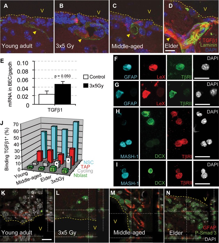
TGF-β/Smad3 signalling increases in the stem cell niche during aging and following irradiation. The data are represented as the mean ± SD The p-value was determined using the Mann–Whitney U-test (*p = 0.020; all of the other p values are given in Supporting Information Table 2). The illustrations are representative of three different experiments, with three mice per group. Scale bars = 10 µm.
A–D. Increasing levels of TGF-β1 were observed in close contact with SVZ microvessels (laminin-positive) during aging and following irradiation.
E. TGF-β1 mRNA expression by qPCR in sorted BECs.
F–I. TβRI and TβRII are abundant on freshly dissociated GFAP+LeX+ and Mash1+ cells.
J. The binding of biotinylated TGF-β1 to neuroblasts (CD24+), NSCs (GLAST+CD24−) and proliferating cells (DNA >2N).
K–N. The phosphorylation of Smad3 was observed in the majority of SVZ cells in irradiated and aged mice, including cells with a GFAP+ type B/NSC phenotype.

