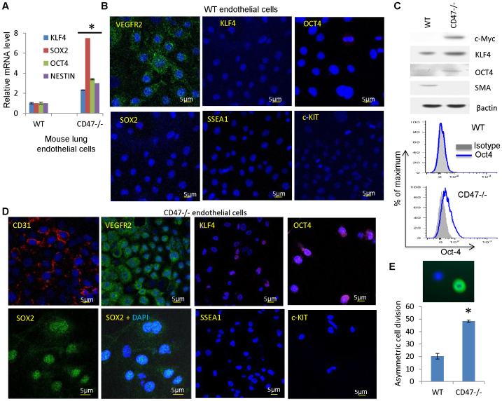
Figure 2. Stem cell and differentiation marker expression in WT and CD47-null endothelial cells.
(A) mRNA expression levels of stem cell transcription factors in WT and CD47 null lung endothelial cells. (B, D) Stem cell and differentiation marker expression in WT (B) and CD47 null mouse endothelial cells (D). The endothelial cells were stained using the indicated antibodies and DAPI (blue). Scale bars = 5 μm. (C) Protein expression of stem cell transcription factors and smooth muscle actin (SMA) assessed by western blotting and flow cytometry (Oct4) in cultured WT or CD47 null endothelial cells in EGM2 medium. (E) Asymmetric cell division frequencies in second passage WT and CD47−/− endothelial cells equilibrium labeled with BrdU and chased for one cell division. Asymmetric division was scored by counting BrdU+(green)/DAPI+ nuclei adjacent to BrdU−/DAPI+ nuclei.

