Figure 6.
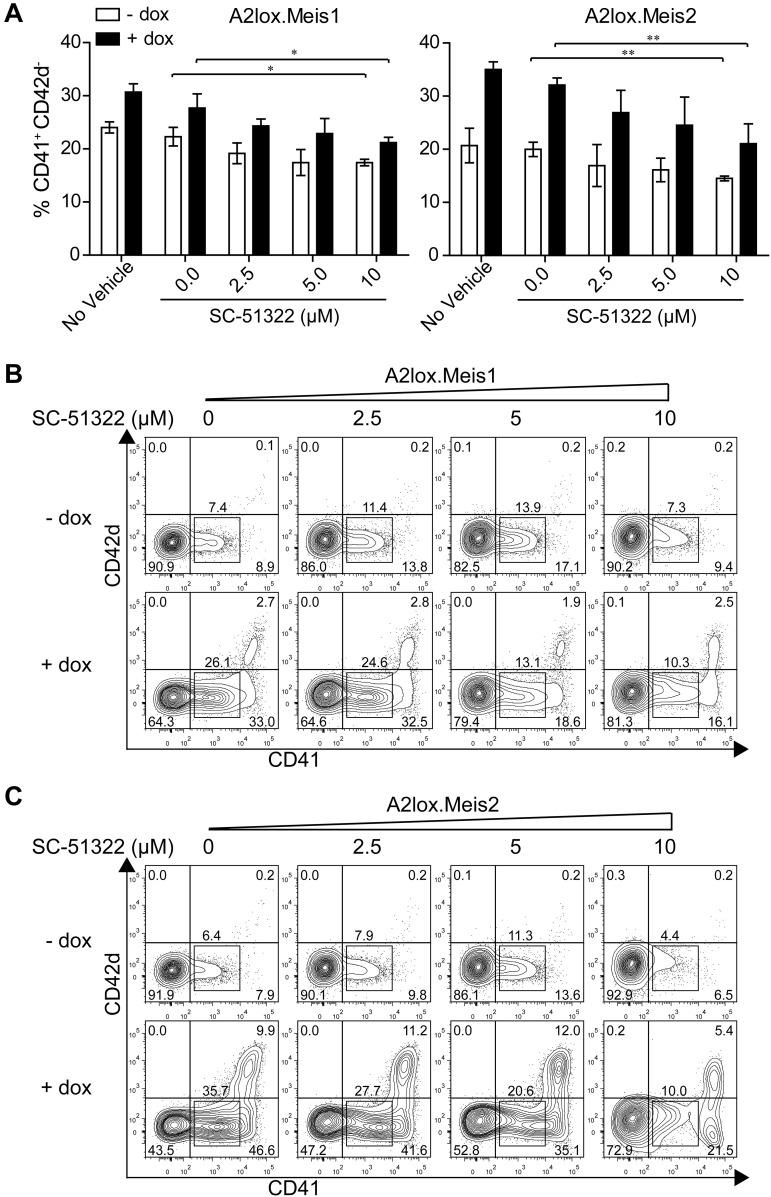
Meis1 and Meis2 induce CD41int hematopoietic progenitors through Ptger3 signaling pathway. (A) A2lox.Meis1 or Meis2 ES cells were differentiated as described in Figure 3A in the presence of the indicated concentration of SC-51322 added on day 6 and replenished daily. On day 9, cells were harvested and analyzed by FACS for expression of CD42d and CD41. Cells are gated as GFP-negative to exclude OP9 cells. Numbers indicate the percentage of CD41+ CD42d− cells developing in the indicated conditions from 3 independent experiments. Bars represent the SD. *P < .05 (paired Student t test). **P < .01 (paired Student t test). (B) A2lox.Meis1 ES cells were differentiated as in Figure 3A with the indicated concentration of SC-51322 added on day 6 and replenished each day until day 12. On day 12, cells were analyzed by FACS for expression of CD42d and CD41. Shown are data for GFP-negative cells to exclude analysis of OP9 cells. Numbers indicate the percentage of cells in the indicated gates. (C) A2lox.Meis2 ES cells were differentiated and analyzed as in panel A.

