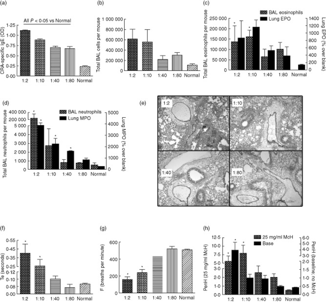
Fig. 1.
Model of mild atopic asthma. Female outbred mice were sensitized and challenged with cockroach allergens for three exposures (days 0, 14 and 21), respiratory physiology measured 3 h post-cockroach allergen (CRA)-challenge and mice killed 5 h post-CRA challenge. Four doses of CRA were tested, each mouse receiving the same dose for each of the three allergen exposures. The x-axis numbers refer to the dilution of the CRA (actual concentrations are found in Table 1). (a) Cockroach allergen-specific immunoglobulin (Ig)E. Allergen-specific IgE displayed a dose-dependent decrease with decreasing allergen concentration. (b) Total bronchoalveolar lavage (BAL) cells. (c) BAL eosinophils and lung eosinophil peroxidase (EPO) activity. (d) BAL neutrophils and lung myeloperoxidase (MPO) activity all display dose-dependent decreases with decreasing allergen concentration. (e) Lung histology depicting inflammatory cell infiltrate ‘i’ and mucin ‘m’. F–H: Pulmonary physiology by whole-body plethysmography (WBP), given for measurements without methacholine. (f) Higher CRA corresponded to lengthened times of expiration, Te. (g) Breathing frequency was depressed significantly with higher CRA concentrations. (h) Airways hyperreactivity represented by enhanced pause (PenH) was exacerbated with higher doses of CRA at baseline and after exposure to 25 mg/ml methacholine (McH). *P < 0·05 versus normal; n = 2–6 per group for one experiment.

