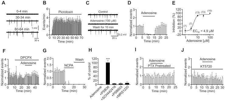
Figure 7. Adenosine-induced depression of seizure activity is mediated by activation of A1 ARs and requires the functions of Gαi proteins and PKA.
A, Seizure events induced by bath application of picrotoxin at the saturated concentration (100 µM) in a rat slice at different times. An extracellular electrode containing ACSF was placed in layer III of the EC to record the seizure events. B, Time course of picrotoxin-induced seizure events (n = 7 slices). C, Seizure events recorded before, during and after the application of adenosine (100 µM). D, Summarized time course of adenosine-induced inhibition of seizure activity (n = 10 slices, p<0.001 vs. baseline, paired t-test). E, Concentration-response curve of adenosine-induced depression of seizure activity. Numbers in the parenthesis are the number of slices recorded from. F, Prior bath application of the A1 AR inhibitor, DPCPX, blocked adenosine-induced depression of seizure events (n = 12 slices, p = 0.89 vs. baseline, paired t-test). G, Bath application of the A1 AR agonist, NCPA, irreversibly suppressed the seizure events (n = 6 slices, p<0.001 vs. baseline, paired t-test). H, Application of antagonists to other ARs except A1 ARs did not block adenosine-induced depression of epileptiform activity (One-way ANOVA followed by Dunnett test, *** p<0.001 vs. adenosine alone). I, Bath application of adenosine failed to depress significantly picrotoxin-induced seizure events in slices pretreated with PTX (n = 8 slices, p = 0.45 vs. baseline, paired t-test). J, Pretreatment of slices with and continuous bath application of the membrane permeable PKA inhibitor, KT5720, blocked adenosine-induced depression of seizure events (n = 8 slices, p = 0.7 vs. baseline, paired t-test).

