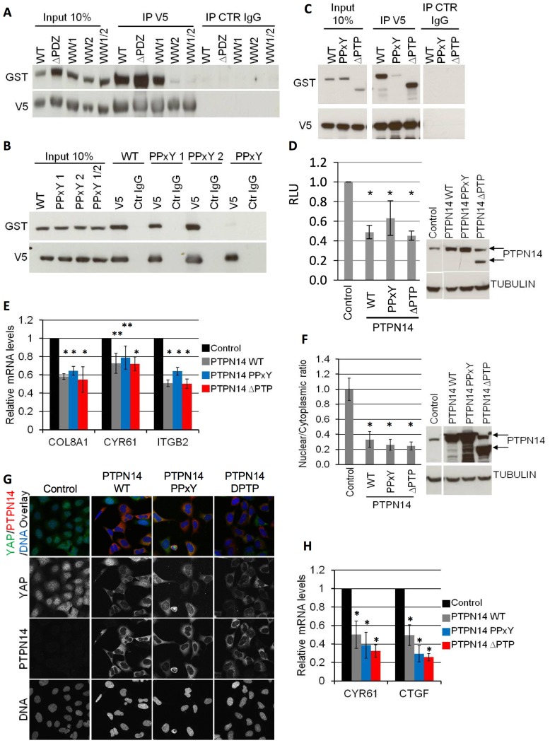
Figure 3. YAP-PTPN14 binding is mediated through the WW domain-PPxY motif interaction.
A) 293A cells were transfected with WT GST-PTPN14 and the indicated V5-YAP constructs (WT and mutants). A V5 IP was carried out and blotted for GST to study the interaction of the various YAP mutants with WT PTPN14. Input lanes were loaded with 10% of the amount of lysate used for each IP and used to compare the expression levels of each construct. B–C) 293A cells were transfected with WT V5-YAP and the indicated GST-PTPN14 constructs (WT and mutants). A V5 IP was carried out and blotted for GST to study the interaction of the various PTPN14 mutants with WT YAP. Input lanes were loaded with 10% of the amount of lysate used for each IP and used to compare the expression levels of each construct. All lanes in (C) are from a single blot and exposure. D) An SF268 cell line stably expressing the YAP-responsive MCAT_Luc reporter was transduced with lentivirus encoding for the indicated dox-inducible PTPN14 expression. Luciferase expression of each cell line was analysed 72 hours post dox induction (left panel). A Resazurin assay was carried out in parallel for each sample and used to normalize the luciferase readings. PTPN14 expression levels achieved for each construct were analysed by Western blot (right panel; arrows indicate the WT PTPN14 protein and the truncated ΔPTP PTPN14 which migrates faster; all lanes from a single blot and exposure). Tubulin serves as loading control. Luciferase results are shown as the average of at least 3 independent experiments ± STDEV. Statistical analysis was carried out with a 2-tailed paired t-test; * p<0.05. E) The mRNA levels of the indicated YAP target genes were assessed in cells from (D) 72 hours post dox induction. Statistical analysis was carried out with a 2-tailed paired t-test; * p<0.001; **p<0.05, F) 293A cells were transduced with lentivirus encoding for the indicated dox-inducible PTPN14 expression. The nuclear/cytoplasmic YAP ratio was quantified at low density after 72 hours of dox induction using a Cellomics automated imager with a conventional microscope, and expressed relative to control (left panel). Results are shown as the average of three experiments ± STDEV. Statistical analysis was carried out with a 2-tailed paired t-test; * p<0.05. For each experiment, the average ratio was calculated from three wells per sample (10 images per well). PTPN14 expression levels were analysed by Western blot (right panel; arrows indicate the WT PTPN14 protein and the truncated ΔPTP PTPN14 which migrates faster; all lanes from a single blot and exposure). Tubulin serves as a loading control G) Confocal microscopy images of 293A cells from (F) generated 72 hours post dox induction. H) The mRNA levels of the indicated YAP target genes were assessed in cells from (F) 72 hours post dox induction. Statistical analysis was carried out with a 2-tailed paired t-test; * p<0.05.

