
Figure 3).
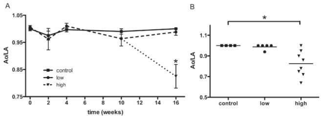
Echocardiographic measurements of the aorta-to-left atrial ratio (Ao/LA). The timecourse of left ventricular (LV) functional response to doxorubicin (DOXO) administration was indirectly inferred by the relationship between the transverse diameters of the aorta and the left atrium (control, n=4; low-dose, n=5; high-dose, n=8). A The Ao/LA is displayed over time, beginning after the completion of the first cycle of DOXO (7.5 mg/kg). There were no significant variations in the ratio between the control and the low-dose groups during the study. However, the high-dose group demonstrated a striking reduction after 16 weeks compared with the control group, indicating LV dysfunction. Data are expressed as mean ± SEM. B Individual results for each animal from tests performed at 16 weeks. Control, low- and high-dose groups are labelled. Control and low-dose groups showed similar results, while nearly all of the animals in the high-dose group exhibited a reduction in the ratio due to the atrial dilation that accompanies LV overload. *P<0.05