
Figure 4).
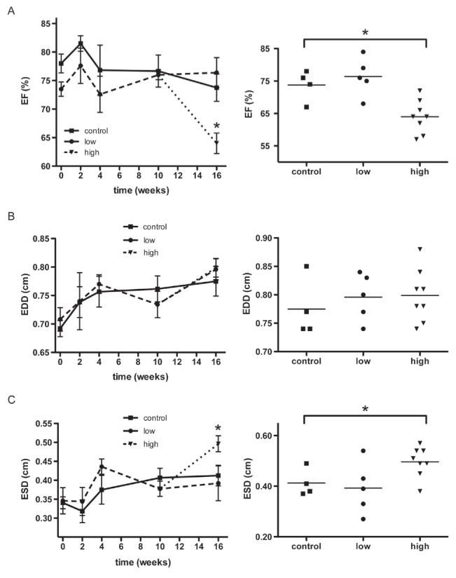
Echocardiographic analysis of left ventricle (LV) systolic performance. A Serial measurements were taken of the ejection fraction (EF) of the LV (left panel). Individual data for the animals from the three groups (control, n=4; low-dose, n=5; and high-dose, n=8) at week 16 is also provided (right panel). The EF of the high-dose group was significantly reduced at 16 weeks compared with the control group. B Serial measurements of the end-diastolic diameter (EDD) of the LV (left panel) and individual data at week 16 (right panel) are shown. There was no significant difference among the three groups. C Serial measurements of the end-systolic diameter (ESD) of the LV (left panel) and individual data at week 16 (right panel) are shown. The high-dose group displayed a significant increase in ESD compared with the control group at 16 weeks. The reduction of the EF in the high-dose group correlated with the increase in ESD in these animals, whereas the EDD did not differ from the control group. These results indicate that the high-dose group experienced LV dysfunction. Data are expressed as mean ± SEM. *P<0.05