Figure 1.
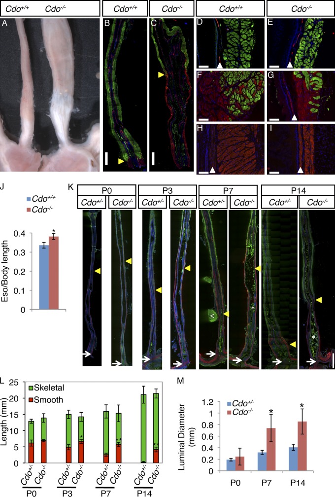
Cdo−/− mice have megaesophagus and display defective proximal-to-distal progression of ME development. (A) Adult Cdo+/+ and Cdo−/− esophagi. (B–I) Longitudinal sections of adult Cdo+/+ and Cdo−/− esophagi stained with antibodies to α-smooth muscle actin (α-SMA; red) and sarcomeric actin (SA; green) for smooth and skeletal muscles, respectively. (B and C) Cdo−/− esophagi have an aberrantly proximally located skeletal–smooth muscle boundary (arrowheads). Only the distal halves of each esophagus are shown (proximal halves have only skeletal muscle). (D and E) The proximal ME of the Cdo−/− esophagus is thinner than the control, but has normal appearing skeletal muscle. The α-SMA+ layer is the muscularis mucosa (arrowheads). (F and G) SMCs are dispersed with skeletal myofibers around the skeletal–smooth muscle boundary of both Cdo+/+ and Cdo−/− esophagi. (H and I) The distal Cdo−/− esophagus has a long, thin extension of smooth muscle instead of the short, broad segment of smooth muscle found in controls. (J) Cdo−/− esophagi are longer than Cdo+/+ esophagi, as normalized to body length (tip of nose to base of tail); values are means ± SD, n = 5. *, P < 0.005. (K) Longitudinal sections of P0, P3, P7, and P14 Cdo+/− and Cdo−/− esophagi were stained with antibodies to α-SMA and SA. The location of the distal-most SA+ cell in each section is denoted by an arrowhead, the LES by an arrow. The white arrowhead in the P7 Cdo+/− section indicates a piece of the SA+ diaphragm still attached to the esophagus; asterisks in the P7 and P14 Cdo−/− sections indicate ingesta (green), which binds antibody nonspecifically. (L) The distance between the distal-most SA+ cell and the LES was measured and is represented by the red portion of the histogram bars. Note that the distance decreases progressively with age in Cdo+/− esophagi, but this fails to occur in Cdo−/− esophagi. Values are means ± SD, n = 3–4. *, P < 0.01; **, P < 0.001; with differences referring to the Cdo+/− and Cdo−/− esophagi at a given age. (M) The luminal diameter of Cdo−/− esophagi is larger than that of Cdo+/− esophagi at P7 and P14, but not at P0. Values are means ± SD, n = 3. *, P < 0.04. Bars: (B, C, and K) 1 mm; (D–I) 0.1 mm.

