Figure 2.
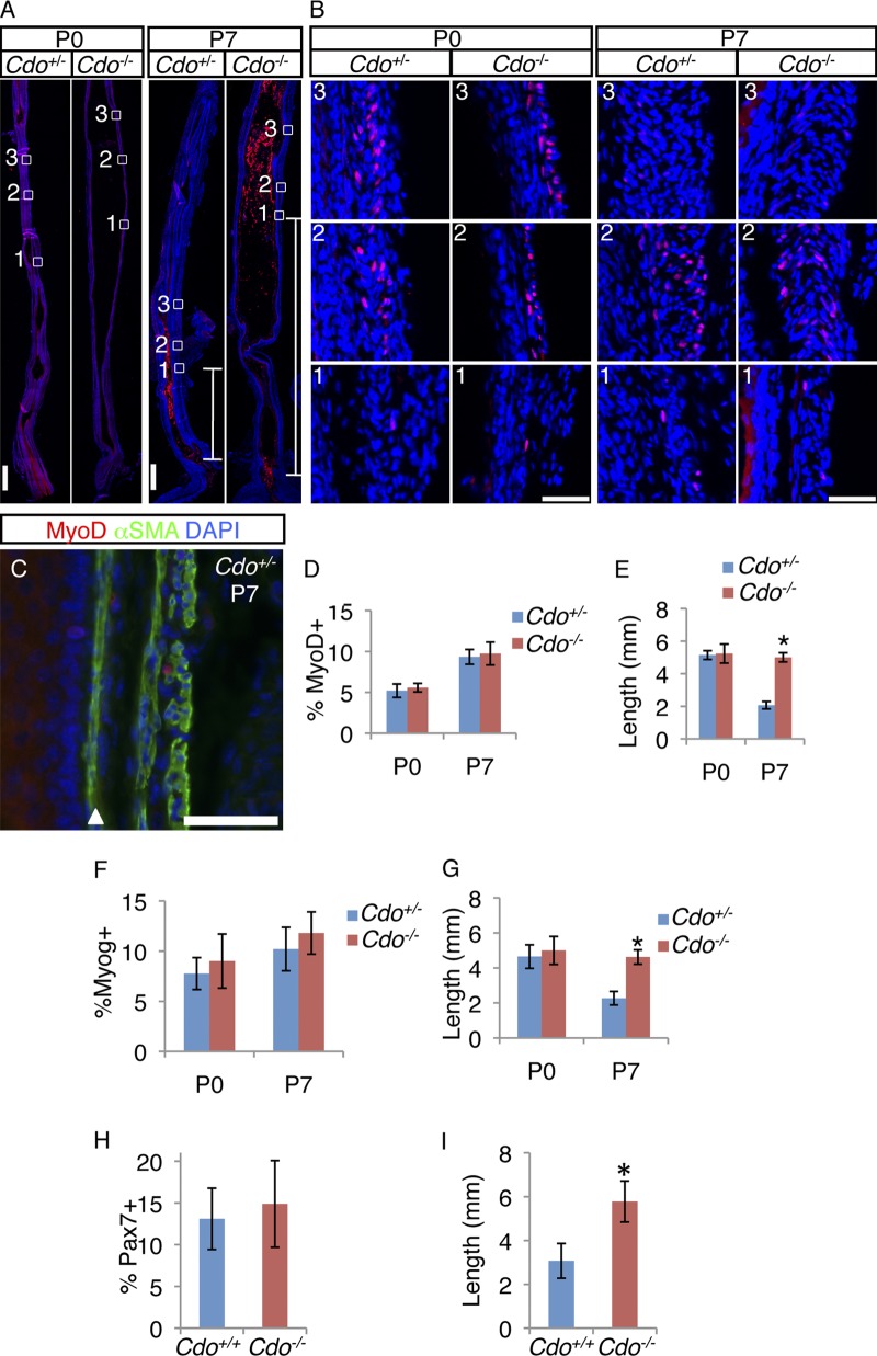
Cdo−/− esophagi have normal numbers of MyoD+, myogenin+, and Pax7+ cells in an aberrantly proximal location. (A) Longitudinal sections of P0 and P7 Cdo+/− and Cdo−/− esophagi were stained with antibodies to MyoD (red). Boxed areas correspond to the distal-most MyoD+ cell (1), the region of the transition zone (TZ) with the greatest number of MyoD+ cells (2), and a more proximal region where, by P7, MyoD+ cell numbers diminish (3). These regions are identified by the presence and numbers of MyoD+ cells and are in different locations along the proximal–distal axis in Cdo+/− and Cdo−/− esophagi. White lines in the P7 micrographs indicate the distance between the distal-most MyoD+ cell and the LES. (B) Higher magnification of boxes 1–3, as indicated. (C) Longitudinal section of P7 Cdo+/− esophagus stained with antibodies to MyoD, α-SMA, and with DAPI. The arrowhead denotes the muscularis mucosa. (D, F, and H) Quantification of MyoD+ (D), myogenin+ (F), and Pax7+ (H) cells within the TZ, measured as a percentage of total DAPI+ cells in the ME. (E, G, and I) Quantification of the distance of between the distal-most MyoD+ (E), myogenin+ (G), and Pax7+ (I) cell and the LES. Values in D–I are means ± SD, n = 3–4. *, P < 0.05. Bars: (A) 0.5 mm; (B and C) 50 µm.

