Figure 3.
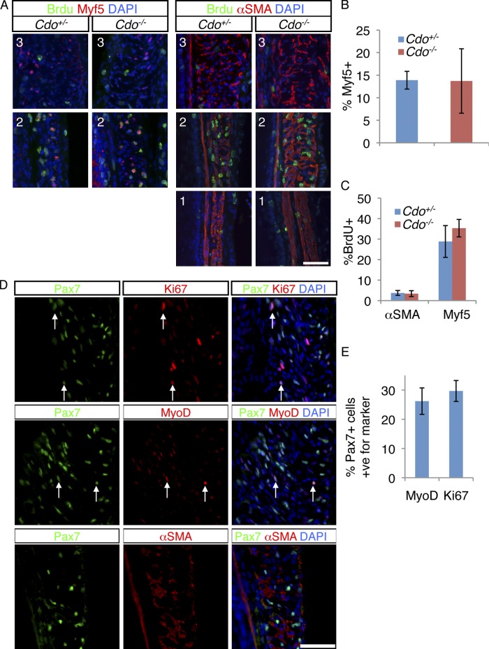
Proliferative skeletal muscle precursor cells are present in the TZ. (A) P7 mice were injected with BrdU and 2 h later longitudinal sections of Cdo+/− and Cdo−/− esophagi were obtained and stained with antibodies to BrdU and Myf5 or α-SMA. The numbers of each panel (1, 2, 3) correspond to the equivalently numbered boxes/panels in Fig. 2. (B) Quantification of Myf5+ cells within the TZ, measured as a percentage of total DAPI+ cells in the ME. (C) Quantification of percentage of Myf5+ and α-SMA+ cells that are also positive for BrdU. Values for B and C are means ± SD, n = 4. (D) Longitudinal sections of P7 Cdo+/+ esophagi were stained with antibodies to Pax7 and either Ki67, MyoD, or α-SMA, and with DAPI. Many Pax7+ cells coexpressed the proliferation marker Ki67, or the muscle determination marker MyoD (arrows in the respective panels), but Pax7+ cells did not express α-SMA. (E) Quantification of the percentage of Pax7+ cells in the TZ that are also either Ki67+ or MyoD+. Values are means ± SD, n = 4.

