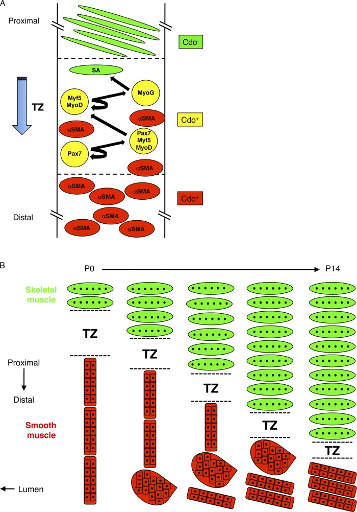
Figure 7.
Models of maturation of the esophageal ME. (A) Model of skeletal myogenesis in the TZ. The TZ contains proliferating skeletal muscle progenitor (Pax7+) cells, muscle progenitor cells in the process of commitment to the skeletal muscle lineage (Pax7+/Myf5+/MyoD+ cells), skeletal myoblasts (Myf5+/MyoD+ cells), and differentiating skeletal myoblasts (Myog+ cells). The TZ moves in a proximal-to-distal manner, leaving SA+ myofibers in its wake. Smooth muscle (α-SMA+) cells are mainly found distal to the TZ where they undergo fascicular reorientation (see panel B). Some SMCs are also found dispersed within the TZ. Cells of the developing skeletal muscle lineage express Cdo, as do SMCs, but mature skeletal myofibers do not. (B) Model for reorientation of SMC fascicles and proximal-to-distal movement of the TZ between P0 and P14. SMCs of the ME are initially grouped into fascicles that have an end-to-end configuration and an orientation parallel to the lumen. They reorganize in a distal-to-proximal manner via a globular intermediate and culminate in a side-by-side configuration with an orientation that is nearly parallel to the lumen; as a consequence, the fascicles ultimately occupy only the most distal portion of the ME. This process clears a path for the TZ, which migrates distally and produces the multinucleated skeletal myofibers that comprise the great majority of the length of the mature ME.

