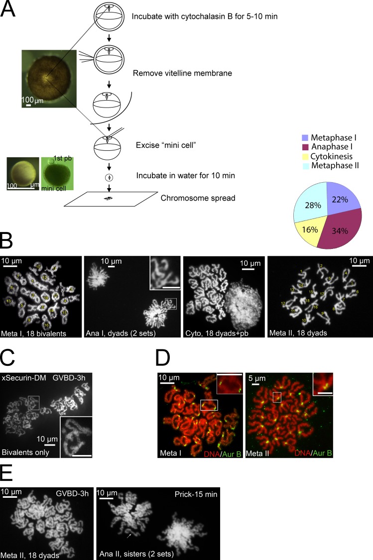
Figure 1.
Karyotyping Xenopus oocytes during meiosis. (A) Schematic illustration of karyotyping Xenopus oocytes: two mini-cells are shown, one without a polar body (left, with overhead light) and the other with a first polar body (pb) still attached (right, photographed with transmission light). (B) Representative images of oocytes at metaphase I, anaphase I, cytokinesis, and metaphase II. The graph summarizes karyotypes of 94 oocytes (>10 experiments) analyzed between 115 and 145 min after GVBD. Numbers are used to facilitate chromosome counting, not to imply chromosome identities. The inset shows two dyads. (C) A representative karyotype image of oocytes injected with xSecurin-DM mRNA at 3 h after GVBD. The inset depicts a single bivalent. (D) Metaphase I (left) and metaphase II (right) chromosome spreads double-stained with Sytox orange and anti–Aur-B. In metaphase I spread, each bivalent (inset) has two pairs of Aur-B foci representing two maternal and two paternal sister centromeres, respectively. In metaphase II spread, each dyad (inset) has two closely associated Aur-B foci representing the two sister centromeres. (E) Metaphase II oocytes before (left) and 15 min after (right) prick activation (n = 7). Note that metaphase II chromosome dyads in these oocytes have more elongated arms than those shortly after polar body emission (see B). Anaphase II spread consists of two sets of sister chromatids (arrow). Bars: (insets) 10 µm.

