
Figure 3.
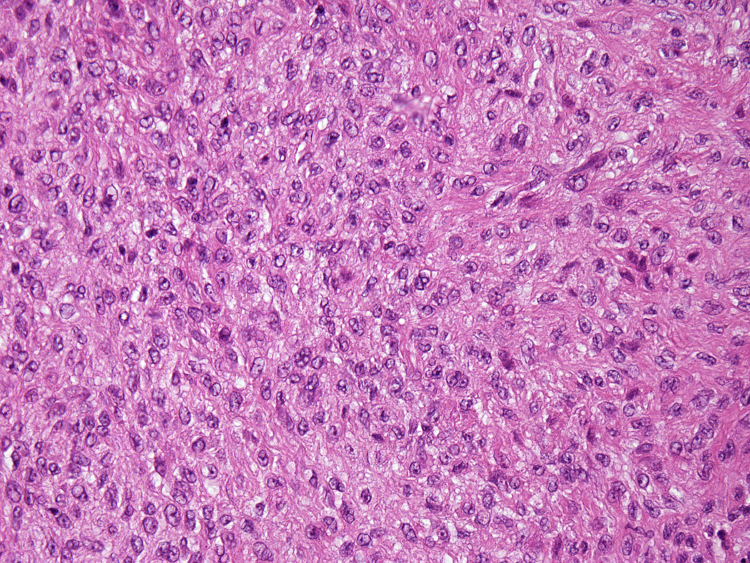
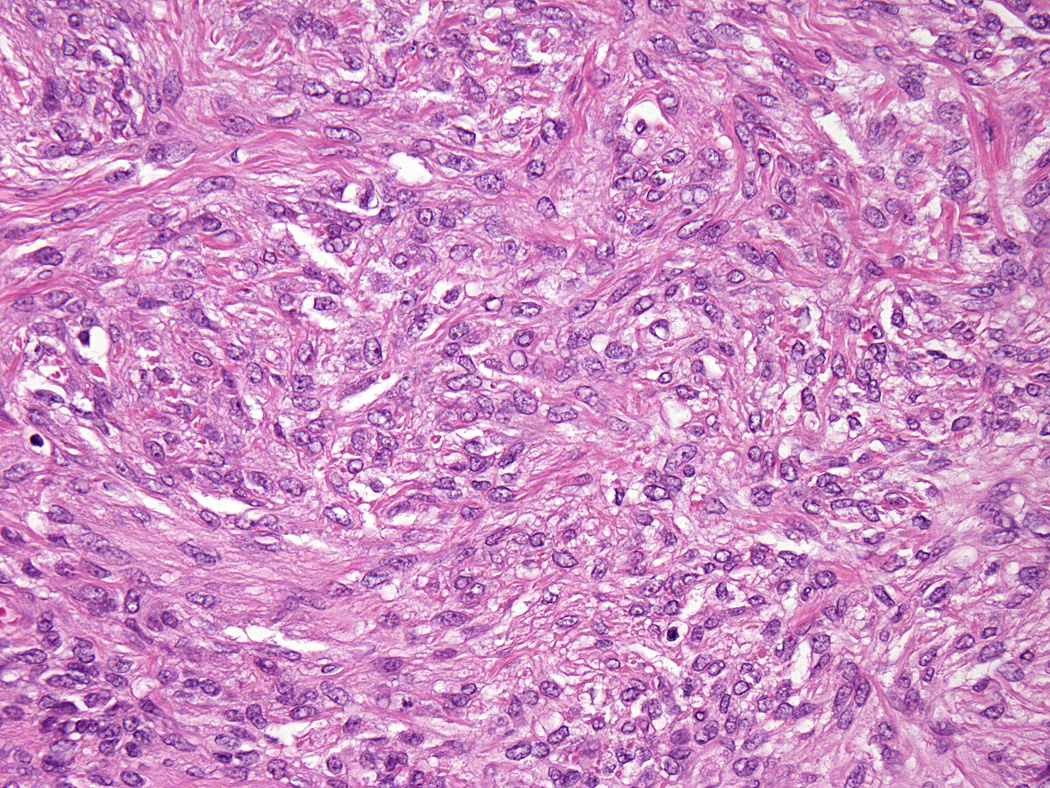
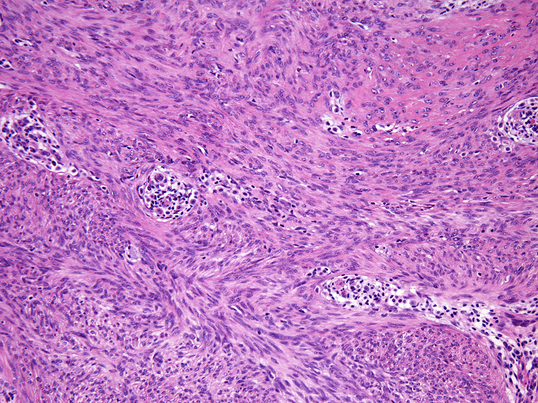
All tumors showed a solid sheet-like growth of uniformly sized ovoid to spindled or histiocytoid cells with palely eosinophilic syncytial cytoplasm (3a); most tumors showed no mitotic activity. Nuclei were vesicular with fine chromatin and smooth nuclear contours and had small or inconspicuous nucleoli, with minimal to no atypia (3b). Focally fascicular growth was observed in only one tumor (3c).