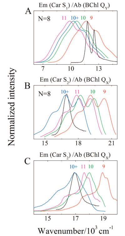
Figure 13.
(A) Spectral overlap between the absorption of the LH2 QX and QY BChl bands from Rb. sphaeroides 2.4.1 and a typical S1 (21Ag−) fluorescence trace taken from 3,4,5,6-tetrahydrospheroidene (N = 8) in petroleum ether shifted to correspond to the spectral origins of the S1 (21Ag−) → S0 (11Ag−) transition of neurosporene (N = 9), spheroidene (N = 10), spheroidenone (N = 10+), and rhodopin glucoside (N = 11). The S1 (21Ag−) energies for the Cars were determined from measurements of the S1 (21Ag−) → S2 (11Bu+) NIR transition energies (Figure 10). (B) Overlap between the absorption spectrum of the LH2 QX BChl band and a typical S2 (11Bu+) fluorescence trace taken from 3,4,5,6-tetrahydrospheroidene (N = 8) in petroleum ether shifted to correspond to the spectral origins of the S2 (11Bu+) → S0 (11Ag−) transition of the Cars in LH2 complexes. (C) Overlap between the absorption spectrum of the LH2 QX BChl band and the S2 (11Bu+) fluorescence taken from neurosporene, spheroidene, spheroidenone, and rhodopin glucoside in n-hexane shifted to correspond to the spectral origins of the S2 (11Bu+) → S0 (11Ag−) transition of the Cars in LH2 complexes.

