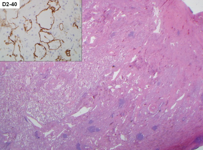
Figure 4).

Low-power view demonstrates a markedly thickened visceral pleura and interlobular septa due to abnormal lymphatic vascular proliferation (hematoxylin and eosin stain, original magnification ×20), as supported by D2-40 immunohistochemical staining (inset, original magnification ×200)
