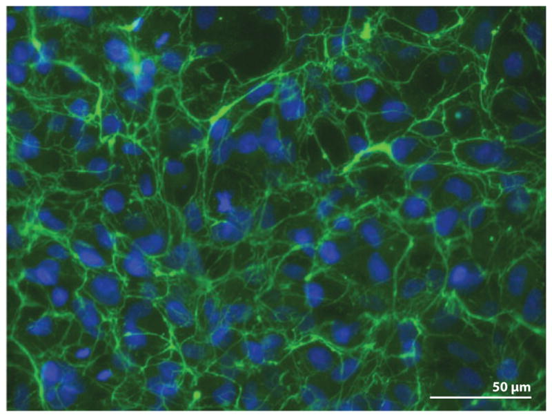
Figure 2.

Fibronectin (FN) fibrillar matrix surrounds cells in culture. HT1080 cells were grown on a glass coverslip for 20 hours in medium supplemented with 0.1 μM dexamethasone and 25 μg ml−1 rat plasma FN as described in Brenner et al. (2000). Cells were fixed and stained with anti-FN monoclonal antibody (IC3) followed by fluorescein-tagged goat antimouse immunoglobulin G. Image shows FN fibrils ( green) around cells with 4′,6-diamidino-2-phenylindole (DAPI)-stained nuclei (blue).
